giulio@gabaldo.com

ESTERS OF ORGANIC ACIDS MFCA (BUTYRIC, CAPRYLIC, CAPRIC, PROPIONIC LAURIC) SHORT AND MEDIUM CHAIN MCFA (MEDIUM CHAIN FATTY ACIDS) ESTERIFIED WITH GLYCEROL IN ANIMAL NUTRITION

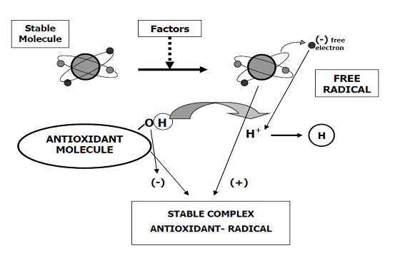
The zootechnical challenge of the next few years that will affect health and productivity in the industrial livestock sector will be to be able to control the oxidative stress of farmed animals by associating at the same time alternative products to the use of antibiotics (antibiotic-free) in order to contain the main problems of the digestive system through the use of natural products and the use of phyto-derivatives with a degree of purity and intracellular concentration obtained “mechanically” without the use of “chemical contaminants” such as solvents with repercussions on animal health , of the environment and of people by combining polyphenols, bioflavonoids, medicinal plant products with anti-inflammatory and medicinal action, MCFA (medium and short chain fatty acids esterified with glycerol) associated with controllers and stimulators of the digestive microbiome such as -pre-postbiotics ( Dr. Giulio Gabaldo – 2018)

BACTERICIDE MECHANISM OF ACTION OF MCFA

(by Ward Dean, MD and Jim English – 2013)

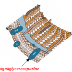
The antibacterial action of the acid depends on the variation of the pH in general. At low pH values ​​they increase the concentration of MCFA (<C4) need to be in the undissociated state to enter the bacterial cell. After entering the cell, the high intracellular pH value leads to their dissociation. The lowering of the internal pH then disrupts the normal metabolism (Ricke, 2003). On the other hand, when the pH rises, it totally loses this ability. Consequently, the antibacterial action is expressed only if the MCFA remains undissociated, that is, in an environment <4.5. When the pH rises ± 7, (second part of the intestine) the MCFA lose this ability and to enter they need the action of the aguaporins which are intrinsic proteins found in the cell membrane wall and which allow the flow of water. in  bidirectional sense. Two families of aquaporins have been identified:
a) Specific aquaporins: they only allow the transport of water. The channel is in fact made up exclusively of amino acids, which only bind water molecules while other ions and molecules do not pass through this channel.
b) Aquaglyceroporins: these also allow the passage of water, but unlike the previous ones, they allow the passage of glycerol and other neutral molecules connected to it.

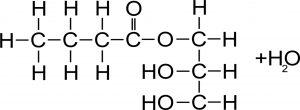
CHEMICAL COMPOSITION OF THE ESTERS OF MCFA ESTERIFIEDWITH GLYCEROL (by SILO – SpA- FI)

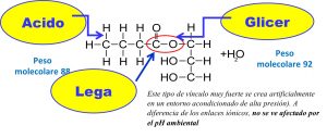
1) Non-esterified MCFA can have an anti-inflammatory and anti-bacterial action only in the 1st part of the intestine (small intestine 2) where the pH is still low (± 4). After that, with the balancing of the pH by the bile to pH ± 7, they become inactive. Increasing the dosages causes loss of appetite and a corrosive action on the intestinal wall.

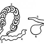
2) MCFA esterified with glycerol, having a stable covalent bond, can “go down” in the lower part of the intestine (fat n ° 3 and 4) in an unaltered form and by means of the aquaglyceroporin mechanism they can “hook” the pathogenic germs (using the glycerol as a transport carrier)

CONCLUSIONS: through the use of esterifieds with MCFA glycerol an effective antibacterial action is obtained without the use of antibiotics ( antibiotic free) on the main intestinal pathogenic families of swine, poultry, horses, etc. (Coli, Clostridia, Salmonella, Brachispira, Rhodococcus, etc.) obtaining at the same time an anti-inflammatory action without harming the positive lactic population of  the intestine


A “sneaky” and “expensive” problem: Coccidiosis and Cryptosporidiosis in calves, little buffalo, lambs and goat kids

A “sneaky” and “expensive” problem: Coccidiosis and Cryptosporidiosis in calves, little buffalo, lambs and goat kids

In young ruminants during weaning (calves, lambs, little capes and buffalettos) diarrhea is considered one of the most important problems affecting the mortality rate of a farm. Among the primary causes we find in the weaning and weaning phase is Coccidiosis (Emeria) and Cryptosporidiosis (or occult spores) whose main forms belong to Cryptosporidium spp. 

The “oocysts” once taken orally by young animals (water, fodder and / or contaminated food) are immediately released into the intestine attacking the intestinal villus where it can persist even for two months. While Cryptosporidiosis can also appear in the first days of life (due to contamination by the mother) Coccidiosis generally strikes during weaning or at the moment of transition between the “milky” and “vegetable” diet.

( by – Management of the Scouring Calf For Irish Farmers, Advisors, Vets -TECHNICAL WORKING GROUP –  Doblin 2011 )

Cryptosporidiosis

 The spores (oocysts) of cryptosporids, once taken orally by calves (water, environment, forages) are active.

Very few are needed to release the sporozoites that colonize the epithelial cells that line the intestinal villi and to manifest the disease.

Contamination occurs orally or from water or food or from a contaminated environment where they can persist from 2 to 6 months. Cryptosporidiosis can also occur at the 3rd-5th day of life but the majority occurs at 2 weeks of age. It is a very contagious disease that manifests itself with diarrhea, anorexia and dehydration even if it rarely leads to the death of calves, as long as they do not involve infections of secondary origin (coli, etc … and / or viral). The course goes from 4 to 18 days and depending on how much and how the villi of the intestine are affected, the appearance of the “diarrhea” (light, dark, etc …) will change.

The trend is strongly influenced by the state of the immune system as well as the expulsion of the oocysts occurs through the feces as early as the 16th day of life. The main form of prophylaxis is:

  1. environmental hygiene, ie preventing the calf from taking on the oocysts
  2. an adequate immune system following a correct intake of good quality colostrum (NUTRICOL IGG> 18%)


Coccidiosis

The infection normally occurs orally (oral-faecal) and spreads with mature oocysts. The fodder and / or water is transmitted directly or through food.

The organism reproduces in the intestine of the host, and thousands of oocysts come to contaminate the environment through feces.

In adequate temperature, humidity and oxygen conditions, the oocysts mature in side of the calf between 3 and 7 days and become able to infect the animal.

Each mature oocyst contains eight “sporozoites”, each of which is able to enter an animal’s intestinal cell after ingesting oocysts and so on. About 70% occurs in the small intestine and where the greatest damage to the intestinal villi occurs

The normal consequences include a loss of absorption surface during the small intestine and a reduced ability to absorb necessary nutrients.


In growing calves coccidiosis has a strong negative impact on the future production performance of cows, buffaloes, goats and dairy sheep.

In all species and in most cases only liquid stools and weight loss are evident, subclinical infections, in fact, are the most expensive since young growing animals (calves, buffaloes, goats and lambs) do not take advantage of the “peak of growth “in the moment of greatest need or in the post-weaning period.


Differential prevention protocol

Protozoa diseases Cryptosporidiosis Coccidiosis
Factor Cryptosporidium spp

Eimeria bovis e Eimeria zuernii, etc.

Incubation   Tendenzialmente prima di 1 mese (5-15 gg)

Fine svezzamento ed l’inizio alimentazione  vegetale

First clinical signs 3-5 gg dopo ingestione oocisti


Durante il periodo di svezzamento

First diarrhea and / or liquid stools 4-5 gg dopo ingestione oocisti
Therapies Symptomatic products (rehydrating and dietetic) Administer feed concentrates and / or feed supplements with “anticoccidials” in the weaning phase
Prevention regulations

1)Use of “potentially effective” colostrum or strengthen the mother’s colostrum (create a company colostrum bank)

2) Complete and radical cleaning of environments and equipment for feeding young animals

3) maximum isolation ofaffected subjects who are nevertheless last fed

4) Registration of affected animals

1) Hygiene and cleaning of environments and equipment for the feeding of young subjects

2) Use of “suitable and good quality” feeds such as cereals for the weaning of young subjects (whole grain and / or extruded barley grains) hay of “excellent” quality grasses, the least contaminated by land as possible

3) If you animals are at risk (in practice almost all farms), administer “coccidiostatic” or medicated feed (with veterinary prescription) and / or specific phytoterapic  products supplements at the beginning of the weaning phase for about 1 month (ex: Alicox granulé)

Disinfection Sunburn and aeration and in addition disinfect with “disinfectants with a strong peroxidant action (7.5% hydrogen peroxide) and / or chloramine T 99% at 0.5% and / or Creolina 0.5 – 1%  


Supplement for farmers


Premix for feed factor


The role and use of antioxidants in nutrition and animal health – Third part

The role and use of antioxidants in nutrition and animal health – Third part

Evaluation systems for the antioxidant capacity of a food

There are practically two systems for evaluating the potential ability to act as an antioxidant for a given food, or to interfere with the metabolic and immune systems of animals:

  • The equivalent antioxidant capacity TEAC (in Trolox) or the equivalent antioxidant capacity of Trolox (TEAC) which measures the antioxidant capacity of a particular substance, compared to the standard, or Trolox (6-hydroxy-2,5,7,8- tetramethylchroman-2-carboxylic acid). It is a water-soluble analogue of Vitamin E. The equivalent antioxidant capacity of Trolox (TEAC) is a Trolox-based antioxidant resistance measurement, measured in units called Trolox micromol ITE, e.g. micromol / 100 g (in vitro examination). Due to the difficulty of measuring the individual antioxidant components of a complex mixture (eg: citrus fruits, olives, blueberries, tomatoes, green tea, etc …), the equivalence of Trolox is , today, used as a reference for the antioxidant capacity of this mixture. The equivalence of Trolox is often measured using the antioxidant capacity of foods (foods rich in polyphenols including those for zootechnical use) as in the ability to reduce plasma iron (FRAP). This test is performed in vitro and only measures the potential antioxidant capacity of the food as a standard value, without verifying its activity at the metabolic level. This method expresses a scale of values called ORAC (Oxigen Radicals Absorbance Capacity).
  • KRL Test (by M. Prost, Kirial – Spirial – Patent M. PROST / SPIRAL – October 2003) KRL test instead provides instead a measure of the status of a subject’s anti-radical global defenses and determines the defensive potential against the free radicals of various products (vitamins, foods, spices, etc …) The test in practice simulates an oxidative attack “type” of red blood cells in a controlled and standardized environment.

Example of ORAC scale

When comparing ORAC (TERAC) data, care must be taken to ensure that the units and the food that is compared are similar. Some evaluations, in fact, will have to be evaluated for ORAC units per gram of dry weight of the fresh whole food (ex: fruit) or of the dry ground or frozen fruit. In each evaluation, different foods may appear with higher ORAC values, therefore it is necessary to compare them with the same parameters (dry, dehydrated and / or whole, etc …)
(eg: in the same way, the large content of water in the watermelon can make it appear as if this fruit were low in ORAC, which is not exact).
Likewise, the typical amount of food used for herbs and spices must be taken into consideration by applying the ORAC scale, but in much lower quantities since we talk about intact whole food concentrates.
Nowadays numerous companies and marketing of food and dietary beverages, animal premixes, etc … have incorrectly capitalized their ORAC rating by promoting the products declared “high in ORAC or TEAC“. As most of these ORAC values are not been objectively validated by independent bodies and / or institutions or subjected to partial revisions for publication in scientific literature, in many cases they remain unconfirmed and not scientifically credible and may mislead users. The Department of Agriculture and Health of the United States (USA) withdrew its reliability in 2012 as biologically invalid, stating that “the data relating to the antioxidant capacity of foods generated by in vitro (test-tube) cannot be extrapolated to the in vivo effects (both for people that animals) and clinical trials to test the benefits of dietary antioxidants have pros unreliable”.

KRL test for resistance to oxidizing factors (by Dr. Michel Prost –SPIRAL – 2003)

KRL test provides a measure of the status of a person’s ( or animal’s )global anti-radical defenses and determines the defensive potential against free radicals of various products (vitamins, antioxidants, foods or feed, etc.)
The test in practice simulates an oxidative attack “type” of red blood cells in a controlled and standardized environment. Under these conditions, the erythrocytes are not affected by other enzyme factors and molecular structures to withstand the oxidative attack until the cell membrane alters to the point of losing their cellular content. The resistance of the erythrocytes tested is therefore expressed by the time taken to release 50% of the hemoglobin content.

The importance of the KRL test is due to provides a measure of the status of a person’s or animal’s global anti-radical defenses and determines the defensive potential against free radicals of various products (vitamins, antioxidants, foods, etc.).The test in practice simulates an oxidative attack “type” of red blood cells in a controlled and standardized environment. Under these conditions, the erythrocytes are not affected by other enzyme factors and molecular structures to withstand the oxidative attack until the cell membrane alters to the point of losing their cellular content. The resistance of the erythrocytes tested is therefore expressed by the time taken to release 50% of the hemoglobin content.

METHOD FOR CARRYING OUT THE KRL TEST (Prost-Spiral patent)

Application of the KRL test

The application of the KIT Radicaux Libres (KRL) was evaluated to evaluate the antioxidant activity of the total blood in pigs (and also in the other animal species). The KRL has also been validated by the FDA – USA and European EFSA and is now considered the most reliable and widely used test in humans to evaluate the effectiveness of natural or pharmaceutical treatments to evaluate natural antioxidant activities (polyphenols and bioflavonoids) or synthetic (vitamin E and vitamin C). The test is recommended as an effective tool for assessing the antioxidant activity of food ingredients in food for pigs (by Rossi R, Pastorelli G, Corino C- Res Vet Sci 2013 Apr).

The principle of RESEDA (Réserves de Défenses Anti Radicalaires – Patent M. PROST / SPIRAL – October 2003)

It has been shown that cells subjected to the metabolic stress of free radicals have the ability to increase their cellular defense systems by accumulating a defense potential against the ones they use in case of need (oxidative stress). This potential changes according to the physiological conditions in which the organism is located and according to the amount of anti free radicals (in practice antioxidants) that the cell has managed to accumulate. This principle (RESEDA) in practice demonstrates the ability of “cellular self-defense” using the accumulated antioxidants (principle patented by Dr. Michel Prost / Spiral October 2003).

SOURCES OF FREE RADICALS (by M.Prost))

CELLULAR DEFENSES AGAINST FREE RADICALS (by M.prost))

CINETIC OF HEMOLYSIS ( by M.Prost )

Results on blood of subjects treated with antioxidants

TEST DONE ON TREATED AND NON-TREATED PIGS WITH ANTIOXIDANTS (by M.prost)

Juridical recognition at French level and validated by the EU of the KRL test

The role and use of antioxidants in nutrition and animal health – Second part

The role and use of antioxidants in nutrition and animal health – Second part

Vitamin E or α-tocopherol

Its activity is mostly focused on the antioxidant action towards to of Carotenes family and Retinol.
The action is attributable to the ability to “break” the chain reactions that produce “free radicals” protagonists of peroxidation.

Since almost all cell membranes are rich in unsaturated fatty acids, the more or less pronounced presence of glutathione-peroxidase (Vitamin E + Selenium) affects the best structural and functional integrity of cell membrane.

The “vitamers” are the tocopherols of which the most active is α-tocopherol. Tocopherols are naturally synthesized from superior vegetables and are mostly found in the form of free alcohols in seeds and leaves.
The metabolic role of Vitamin as an antioxidant factor, in the prevention of oxidation of polyunsaturated fatty acids, a key phenomenon in the development of the fat peroxidation process.

The action of “free radicals” develops through chain reactions that continue the process therefore Vitamin E is:
a) capable of blocking this phenomenon by donating a hydrogen atom (oxidation) to peroxy lipidic radicals, thus making them less reactive and effectively blocking lipid peroxidation.
b) it was shown that tocopherol can interfere with the activity of certain calcium / phospholipid-dependent kinases or protein kinases C (PKCs) interacting directly acting as an anti-proliferative on tumor cells. The action of tocopherol as such or as an organic derivative (succinate) on the growth of malignant cells has been proven for some time.

A group of Italian researchers from the University of Ann Harbor in Michigan has discovered how tocopherol can exert direct effects on gene expression. The studies of this group led to the discovery of a cellular cytoplasm protein able to bind tocopherol (Tocopheryl-Activated Protein-1; TAP-1) and to program the expression of specific genes.
The coordinated action of these genes would allow specific responses at the cardiovascular, immunological, nervous and cartilaginous levels.

The actions and mechanisms by which vitamin E acts in the body were almost completely obscure until a decade ago.
This reaction called “redox” transforms vitamin E into an α-tocoploxylic radical which is very stable, thanks to the development of resonance phenomena, and which can react with Vitamin C or glutathione or Co-enzyme Q10 to reform the α-tocopherol. Since the development of lipid peroxidation can determine profound alterations of cell membranes, we understand why vitamin E is recognized as having an important role in keeping these structures undamaged.

This is also verified by the fact that red blood cells (erythrocytes), which are particularly subjected to oxidative stress, are affected quite early by vitamin E deficiencies, becoming more sensitive to haemolysis. (test by KRL- M.Prost – Spiral). Vitamin E also regulates the activity of two enzymes (lipoxygenesis and cyclooxygenesis) involved in the formation of compounds capable of mediating platelet aggregation phenomena which are accentuated by the lack of the vitamin (prostanoids)

Bioflavonoids and polyphenols and their antioxidant role

They constitute a family of a few thousand (more than 5000) of natural and semi-natural organic molecules widely found in the plant kingdom. It is a very large group of derivatives of the secondary metabolism of plants and is characterized, as the name indicates, by the presence of multiple associated phenolic groups in more or less complex structures generally of high molecular weight such as highly polymerized phenolic acids such as tannins (not soluble) The number and characteristics of these phenolic structures underline the unique physical, chemical, and biological (metabolic, toxic, therapeutic, etc.) properties of particular members of the polyphenol class. The number and characteristics of these phenolic structures underline the unique.

The role and use of antioxidants in nutrition and animal health – First part

The role and use of antioxidants in nutrition and animal health – First part

What are oxidizing substances or ROS (Reactive Oxygen Species)?

In chemistry it is said that a chemical element undergoes oxidation when it undergoes an electron subtraction, which translates into an increase in its oxidation number. This subtraction of electrons can take place by another element, which thus undergoes the complementary reduction process. Most oxidation reactions involve the development of energy in the form of heat and electricity. Substances that have the ability to oxidize other substances are known as oxidizing agents or ROS.

They subtract electrons from other substances and in practice accept electrons. Oxidizers are generally chemical substances that possess elements with a high number of oxidation, for example hydrogen peroxide, permanganate or highly electronegative substances such as oxygen (eg: air), flower, chlorine (eg: sea salt) or bromine, capable of removing one or more electrons from other substances.


Oxidation

Simple classic examples: Piece of oxidized metal (corroded) – Corrosion

Oxidation is a chemical reaction that transfers electrons from a substance to an oxidizer.
Oxidation reactions produce free radicals or ROS, which are responsible for initiating a chain reaction that damages cells. Antioxidants terminate these chain reactions by intervening on intermediate radicals and inhibiting other oxidation reactions by oxidizing themselves.

Oxidative stress

Oxidative stress is a pathological condition caused by the breakdown of physiological equilibrium, in a living organism (vegetable, animal or human), between the production and elimination, by antioxidant defense systems from oxidizing substances.
All life forms maintain an “antioxidant reducing environment” (antioxidant stock) within their cells. In the REDOX cellular environment (with the term redox or redox from the English REDduction, reduction and OXidation, oxidation) all those chemical reactions take place in which the oxidation number of the atoms changes (ie all the reactions in which there is an exchange of electrons from one chemical structure to another) it is preserved by enzymes that maintain the reduced state through a constant input of metabolic energy.
Possible alterations of the normal REDOX state can have toxic effects for the production of peroxides and free radicals that damage all the components of the cell, including proteins, fats and DNA intervening negatively in the systems of self-defense (immunodepression) and in the health of the organis.


Cellular oxidative stress

Oxidative stress, on the part of free radicals and how, these oxidative processes, can cause significant oxidation at the level of the cell membrane and destroy DNA. Today it is possible to evaluate them by means of tests that help us assess the state of health of the organism , the inflammatory state and the onset of some diseases (eg: in humans pathologies such as diabetes, Alzheimer’s and cardiovascular diseases, but also in animals as in pigs with the onset of “very aggressive viral and bacterial forms that are not very sensitive to normal drugs, such as PRRS, etc … or even, not less important, lack of productive and qualitative performances).
Today it is possible to measure both the production of free radicals and the body’s ability to react to oxidative stress through the antioxidant barrier that includes both endogenous antioxidants (complex enzyme systems) and exogenous ones (ie those that are taken through nutrition), and also antioxidant power of a particular functional food (KRL test on red blood cells).


Classic example “on the apple” of damage from free radicals at the cellular level


Definition of antioxidants

Antioxidants are natural and non-natural chemical substances (molecules, ions, radicals) or physical agents that slow down or prevent the oxidation of other substances as a result. The antioxidants are chemically defined of involve as reducing agents (such as thiols and polyphenols) as the chemicals involved in to reactions in thr oxide-reducing. Although oxidation reactions are vital for life, they can be just as harmful; therefore, both plants and animals maintain multiple types of antioxidants as complex self-defense systems.


Antioxidants can be ….

  • Primary:
    1) When they prevent the production of “species” of radicals
    2) When “grappling ” on the transition metals
  • Secondary: when they react with the newly formed radicals and convert them into non-reactive forms by interrupting the chain reaction and therefore can be:
  1. Endogenus: quando sono sintetizzati dall’organismo stesso( enzimatici cellulari) ed a seconda della loro azione posso essere
    a) 1) Enzyme type cellular, as:
    – SOD (superoxide dismutase), catalase and glutathio-peroxidase (it is the main cellular antioxidant that maintains low O2 level and works in conjunction with Catalase and Glutathione Peroxidase (GSH-Px —-> Vit E + Se) it is the main “detoxifier” of the cells:


    – 2. The Catalase (CAT): 2 H2 O2 ——> 2 H2O + O2
    b) Type Protein as SH and metallic sequestering agents (Fe, Cu,)

  2. Exsogenus:
    – Vitaminics: Vitamin C and Vitamin E and Carotenoids (as provitamin A)
    – Polyphenols and Bio-flavonoids


How do antioxidants work?

The oxidation process is a chemical reaction that transfers electrons from a substance to an oxidizer

The intoxication subacute of ammoniacal nitrogen in the nutrition of the dairy cow of high production

The intoxication subacute of ammoniacal nitrogen in the nutrition of the dairy cow of high production

The nitrogen metabolism in dairy cows:
(by M. Wattiaux – Babcock Institute for International Dairy Research and Development University of Wisconsin – 2014)

The nitrogen metabolism in dairy cows:

The ruminal pH change caused by a sudden and / or sharp increase in rumen soluble nitrogen (protein imbalance), resulting in:

  • one excessive ingestion of highly soluble fresh protein fodder (ex: excessive doses of green pasture rich in soluble nitrogen such as alfalfa and / or clover, etc …)
  • the use of high doses of silage with a high content of NH3 in free form in the ration, etc … causes both a soluble nitrogen poisoning in the rumen which can also be followed by an acute alkalosis (but only on some subjects) and more as one increase in ammoniacal nitrogen at a ruminal habitat followed by one metabolic level of NH3 (subacute form) which then, in practice, is also the most damaging for economic purposes as it covers the whole herd


Effects on animal health and production:

This situation at rumen level creates the ideal conditions for the development of the Allisonella histaminiformans, a ubiquitous microorganism of the rumen producing histamine that results in:

  • the immediate inflammation of the rumen papillaes (due to increased circulating cytokines) and consequent reduction in the assimilation of AGVs (Volatile Fatty Acids) followed by the activation of diseases affecting:
    • udder (increase in the infimmatory state in the breast and consequently more CSS and mastitis)
    • feet (formation of thrombus at amatic level >> interdigital phlegmon >> laminitis)
    • ovaries (ovarian cysts)
  • in some animals the acute form (acute alkalosis) may also occur with diseases affecting:
    • liver (hepatic steatosis)
    • of the kidneys (nephritis)
      and in severe forms
    • CNS (neuroplegic symptoms with ataxia, impaired gait etc … and in some cases coma and death of subject Struck)

Actions to be taken on the ration for the subacute forms to improve the health, reproduction and production of cows:

  • Elimination and / or reduction of the toxic nitrogen food source ( green grass, grass silage, urea, etc …) and
    add the following mix to the ration
  • mix of sodium propionate 100 g / head / day + 20 g. / head/ days of Micronil ® (ProbioactiFAP®) + ANTIGRIP FEED (NUTRIVIT-COFATHIM phytotherapic with an anti-inflammatory action ) 50g./head/days This mix must be administered until the end of the use of the “toxic” ( ex: grass silage ) feed source in the diet and continue for at least another 10 days and continue for at least another 10 days.

BIBLIOGRAPHICAL REFERENCES
– Anon. Third External Review Draft of Air Quality Criteria for Particulate Matter (April, 2002). Volume I, II. EPA. United States Department of Environmental Protection Agency. www.epa
– Bach A., Calsamiglia, S. and Stern, M.D. 2005. Nitrogen Metabolism in the Rumen J. Dairy Sci., 88: 9 – 21 Baker, L.D., J.D. Ferguson, and C.F. Ramberg. Kinetic analysis for urea transport from plasma to milk in dairy cows. J. Dairy Sci. 75 (Supplement 1):181, 1992.
– Baker, JL, 2001. Limitations of improved nitrogen management to reduced nitrate leaching and increase use efficiency. Optimizing Nitrogen Management in Food andEnergy Production and Environmental Protection: Proceedings of the 2 nd International Nitrogen Conference on Science and Policy. The Scientific World 1(S2), 1016.
– Cowling, E., J. Galloway, C. Furiness, M. Barber, T. Bresser, K. Cassman, J.W. Erisman, R.Haeuber, B. Howarth, J. Melillo, W. Moomaw, A. Mosier, K. Sanders, S. Seitzinger, S.Smeulders, R. Socolow, D. Walters, F. West, and Z. Zhu. 2001. Optimizing nitrogen management in food and energy production and environmental protection: Summary Statement from the Second International Nitrogen Conference. TheScientificWorld 1(S2): 19. DePeters, E.J. and J.D. Ferguson. 1992. Nonprotein nitrogen and protein distribution in the milk of cows. J. Dairy Sci. 75:31923209.
– Dou, Z., D.T. Galligan, C.F. Ramberg, Jr., C. Meadows, and J.D. Ferguson. 2001. A survey of dairy farming in Pennsylvania: Nutrient management practices and implications. J. Dairy Sci. 84:966973.
– Ferguson, J.D., Z. Dou, and C.F. Ramberg, Jr. 2001. An assessment of ammonia emissions from dairy facilities in Pennsylvania. TheScientificWorld 1(S2): 348355. Erickson, G.E. and T.J. Klopfenstein. 2001. Nutritional methods to decrease N losses from opendirt feedlots in Nebraska. TheScientificWorld 1(S2): 836843.
– Ganong, W.F. Review of Medical Physiology. Nineteenth edition . Co 1999. Appleton and Lange a Simon & Schuster Company. Stamford, Ct. 069120041.
– Hof, G., M.D. Vervoorn, P.L. Lenaers, and S. Tamminga. 1997. Milk urea nitrogen as a tool to monitor the protein nutrition of dairy cows. J. Dairy Sci. 80:33333340.
– Huhtanen, P. 1998. Supply of nutrients and productive responses in dairy cows given diets based on restrictively fermented silage. Agric. Food Sci. Finl. 7:219–250
– Jarvis, S.C., D.J. Hatch and D.H. Roberts. 1989a. The effects of grassland management on nitrogen losses from grazed swards through ammonia volatilization; the relationship to excretal N returns from cattle. J. agric. Sci. Camb. 112:205216.
– Jarvis, S.C., D.J. Hatch and D.R. Lockyer. 1989b. Ammonia fluxes from grazed grassland: annual losses from cattle production systems and their relation to nitrogen inputs. J. agric. Sci. Camb.113:99108.
– Jonker, J.S., R.A. Kohn, and R.A. Erdman. 1998. Using milk urea nitrogen to predict nitrogen excretion and utilization efficiency in lactating dairy cows. J. Dairy Sci. 81:26812692.Muck, R.E. and B.K. Richards. 1983. Losses of manurial N in freestall barns. Agric. Wastes 7:6579.
– Muck, R.E. 1982. Urease activity in bovine feces. J. Dairy Sci. 65:21572163.
– Muck, R.E. and F.G. Herndon. 1985. Hydrated lime to reduce manorial nitrogen losses in dairy barns. Transactions of ASAE 28:201208.
– NRC. 2001. Nutrient Requirements of Dairy Cattle. Seventh Revised Edition. National Academy Press. Washington D.C. NRC. 1996. Nutrient Requirements of Beef Cattle. Seventh Revised Edition. National AcademyPress. Washington D.C.
– Roseler, D.K., J.D. Ferguson, C.J. Sniffen and J. Herrema. 1993. Dietary protein degradability effects on plasma and milk urea nitrogen and milk nonprotein nitrogen in Holstein cows. J. Dairy Sci. 76:525534.
– Scholefield, D., D.R. Lockyer, D.C. Whitehead, and K.C. Tyson. 1991. A model to predict transformations and losses of nitrogen in UK pastures grazed by beef cattle. Plant and Soil132:165171.
– Smits, M.C.J., H. Valk, A. Elzing, and A. Keen. 1995. Effect of protein nutrition on ammonia emission from a cubicle house for dairy cattle. Live. Prod. Sci. 44:147156.
– Voorburg, J.H. and W. Kroodsman. 1992. Volatile emissions of housing systems for cattle.Livestock Prod. Sci. 31:5770.
– Wattiaux , M.A – Protein Metabolism in Dairy Cows – Babcock Institute for International Dairy Research and Development – University of Wisconsin-Madison -2014
– Wilkerson, V.A., D.R. Mertens, and D.P. Casper. 1997. Prediction of excretion of manure and nitrogen by Holstein dairy cattle. J. Dairy Sci. 80:31933204.
– Van Horn HH. 1991;Managing Dairy Manure Resources to aviod Environmental pollution. J Dairy Sci 77:2008-1994.
– Van Horn HH. Balancing nutrients, manure use reduces pollution. Feedstuffs. The Miller Publishing Co. 1992; 64(Oct. 26, 1992). 11-23. Minnetonka, MN.
– Vanfaassen HG, Lebbink G. 1994;Organic matter and nitrogen dynamics in conventional versus integrated arable farming. Agr Ecosyst Environ 51:209-26.
– Vanhorn HH, Wilkie AC, Powers WJ, Nordstedt RA. 1994;Components of Dairy Manure Management Systems. J Dairy Sci 77:2008-30. Webb J, Archer JR. ; Dewi IA, Axford RFE, Marai IFM, Omed H, editors.Pollution in Livestock Production Systems. Oxon, UK: CAB International, 1994; 11,
– Pollution of Soils and Watercourses by Wastes from Livestock Production Systems. p. 189-204.
– Young CE, Crowder BM, Shortle JS, Alwang JR. 1985;Nutrient Management on Dairy Farms in Southestern Pennsylvania. J Soil Water Conserv 40:443-445.